X-Ray
The X-Ray was first discovered in 1895 by Wilhelm Rontgen and soon after, physicians and physicists began to use X-Rays on patients to investigate the skeleton, lungs and other organs. This was the birth of modern-day radiology.
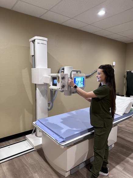
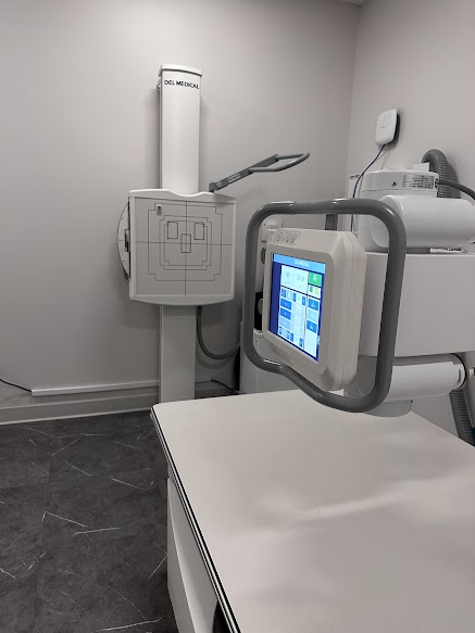
Today, X-Rays use a far safer form of external radiation to produce images of the body, organs, and other internal structures for diagnostic purposes. X-Ray beams pass through body structures onto specially treated plates (similar to camera film) or digital media, and a type of picture similar to a “negative image” is produced. The more solid a structure is, the whiter it appears on the film.
Ahwatukee
4530 E Ray Rd, #160, Phoenix, AZ 85044 | (480) 893-1004
Apache Junction
1840 W Apache Trail, Apache Junction, AZ 85120 | (480) 288-6400
X-Ray Procedure
When the body undergoes an X-Ray, different parts of the body allow varying amounts of the X-Ray beams to pass through. The soft tissues (such as blood, skin, fat and muscle) allow most of the X-Ray to pass through and appear dark gray on the film or digital media. A bone or tumor, which is more dense than soft tissue, allows few of the X-Ray beams to pass through and appears white on the X-Ray. When a break in a bone has occurred, the X-Ray beam passes through the broken area and appears as a dark line in the white bone.
X-Ray technology is used in other types of diagnostic procedures, such as computed tomography (CT) scans and fluoroscopy.
Radiation during pregnancy may lead to birth defects. Always tell your radiologist or doctor if you suspect you may be pregnant.
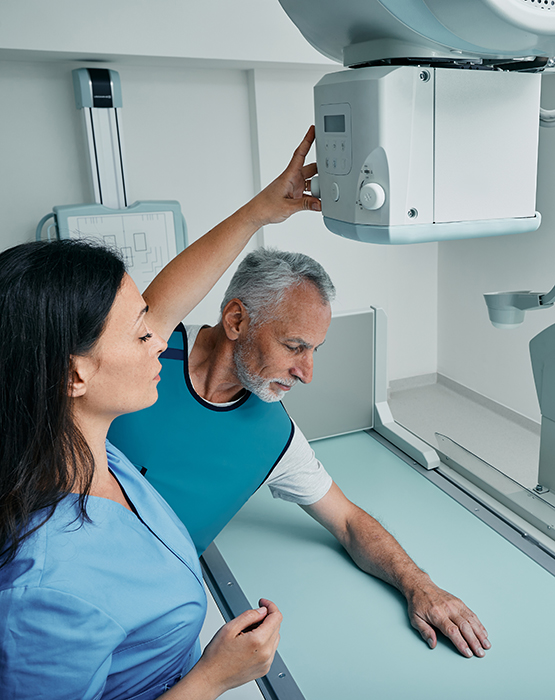
X-rays and general radiography play a very important role in helping physicians make their diagnostic decisions for treatment. X-ray finding often suggests to the radiologist that further diagnostic imaging procedures will be required. The procedure usually takes between 10 to 30 minutes depending on the type of exam requested. Some patients may be asked to change into a gown if their clothing has zippers or buttons that are in the view.


