Our Services
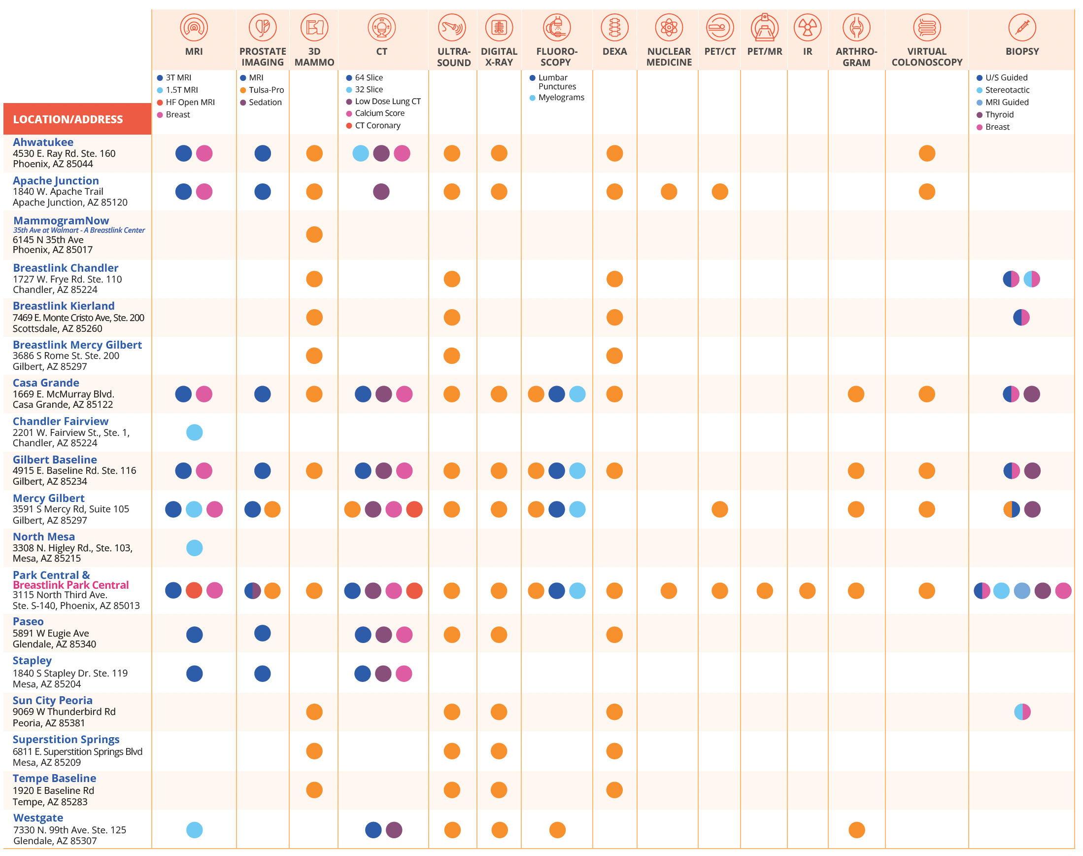
At Arizona Diagnostic Radiology, we offer a comprehensive range of advanced imaging services, utilizing the latest technologies and state-of-the-art equipment to ensure precise and reliable diagnostics.

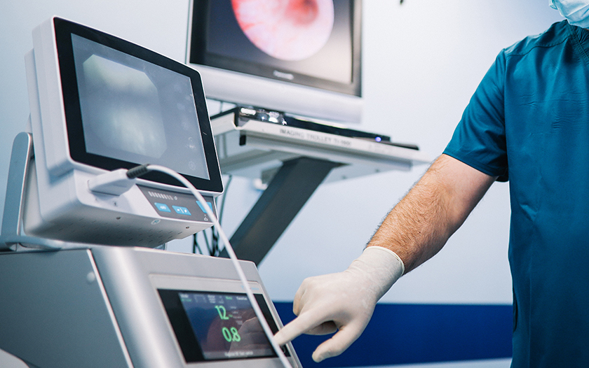
TULSA Procedure
Thousands of men are diagnosed with prostate cancer and enlarged prostate (BPH) each year. The TULSA Procedure offers these patients an incision-free outpatient solution for prostate disease with no radiation, minimal to no pain, a speedy recovery, and a decreased risk of the side effects common to most invasive prostate surgeries.

High-Risk Breast Program
There are a number of benefits to patients who discover their risk for hereditary gene mutations for cancer. This can provide patients the opportunity to understand and, in many cases, manage cancer risk and make decisions about personalized screening and prevention. Genetic testing also allows family members to learn about their own cancer risks.
Our High-Risk Breast Program can identify patients who are at an elevated risk for developing breast cancer, based on family history or inherited genetic mutations.

Low-Dose CT Lung Cancer Screening
Low-dose computed tomography (LDCT) screening for lung cancer is a specialized, painless test that uses low doses of radiation in order to find lung cancer before it is symptomatic and before it has spread.
Lung cancer remains the leading cause of cancer death in the United States but the key to surviving cancer is finding it in its earliest stage, when treatment is most likely to be successful. Several prominent groups, including the American Cancer Society, recommend anyone at high risk for lung cancer to begin screening with low-dose chest CT at the age of 50.
MRI/MRA
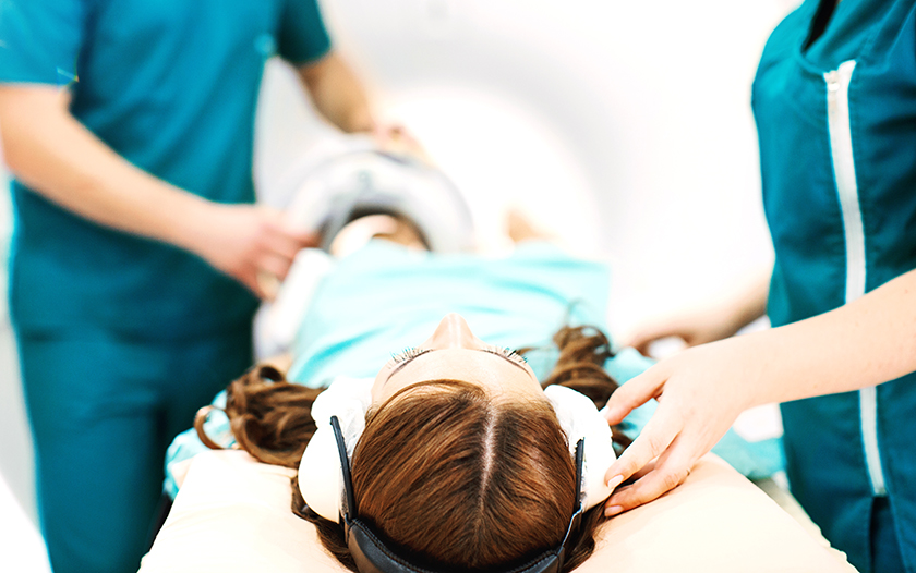
MRI and MRA are both types of scans used to create detailed images of internal body structures such as organs, tissues and bones. The MRI machine creates a magnetic field which bounces radio waves through the body which work to map the scanned section being imaged.
Both MRI (magnetic resonance imaging) and MRA (magnetic resonance angiography) are noninvasive and painless diagnostic tools.
CT Scan
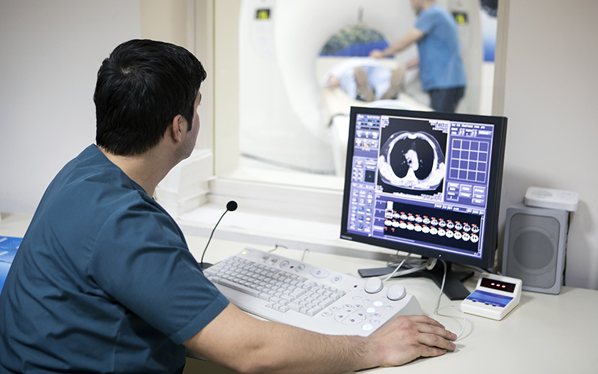
A CT scan is a noninvasive type of X-Ray that creates multiple, cross-sectional images of the body which allows for a detailed examination of internal structures. This exam helps radiologists and physicians diagnose medical conditions that may not be visible on other types of imaging technology.

Dexa Scan
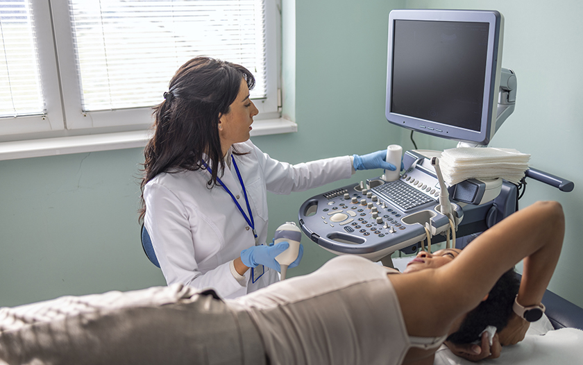
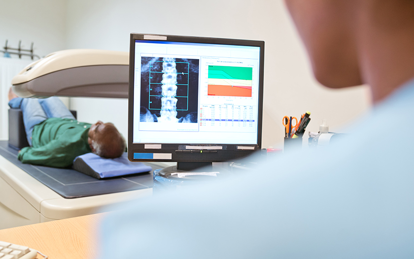
A DEXA scan, also known as a bone density scan, is a painless, noninvasive X-Ray procedure that measures bone strength and thickness. It is most often used to diagnose osteoporosis, a condition that weakens bones and increases the risk of fractures. DEXA scans can also be used to assess the effectiveness of osteoporosis treatments and to predict the likelihood of future fractures.
Nuclear Medicine
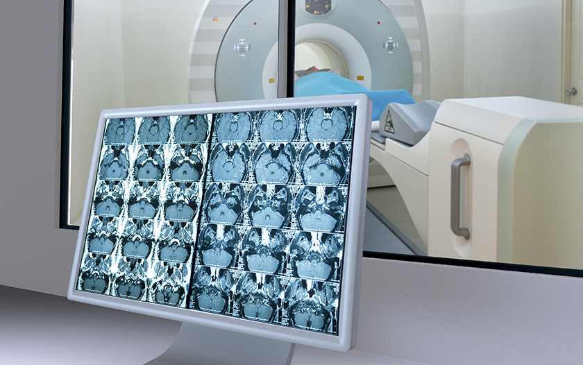
Nuclear medicine is a specialized area of radiology that uses very small amounts of a radioactive substance for the diagnosis and treatment of various conditions, including cancer. Nuclear imaging shows organ and tissue structure more clearly, as well as blood circulation, and how well organs or tissue are working.

PET Scan
Positron emission tomography (PET) scans detect early signs of cancer, heart disease and brain conditions. The procedure requires an injection of a small amount of a radioactive tracer which allows the scanner to make detailed, computerized pictures of areas inside the body.
A PET scan measures important body functions, such as blood flow, oxygen use, and sugar (glucose) metabolism, to help doctors evaluate how well organs and tissues are functioning.

Breast Biopsy
A breast biopsy is a procedure that removes a small amount of breast tissue for examination under a microscope in order to check for cancer or other abnormal cells. A breast biopsy is typically recommended if you have a suspicious area in your breast, such as a lump, or if an abnormality is seen on an imaging test, for instance, a mammogram or breast ultrasound.

Breast MRI
Breast MRI (magnetic resonance imaging) is a powerful diagnostic tool for evaluating breast health. Breast MRI uses radio waves and a strong magnetic field to create detailed images of breast tissue, which can aid in detecting and characterizing abnormalities. Breast MRI is used in conjunction with mammography to enhance breast cancer diagnosis.
Prostate MRI
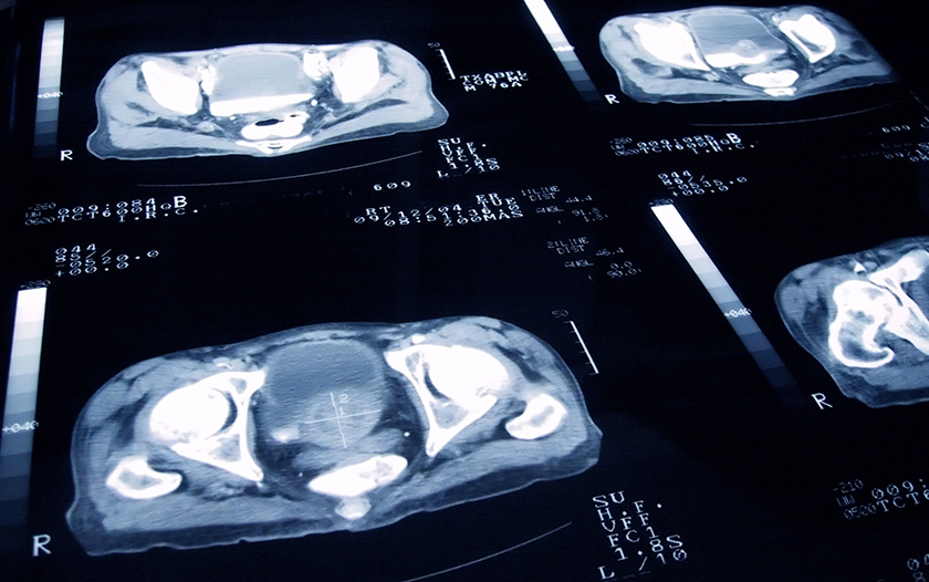
Prostate MRI (magnetic resonance imaging) is an advanced imaging technique that uses a large magnet, radio waves and a computer to produce detailed pictures of the structures inside the prostate gland. It is most often used to assess prostate cancer and evaluate whether it has spread outside the gland. It may also be used to help plan radiotherapy for prostate cancer, assess whether prostate cancer has returned after treatment, and diagnose infection, benign prostatic hyperplasia (BPH) or congenital abnormalities.
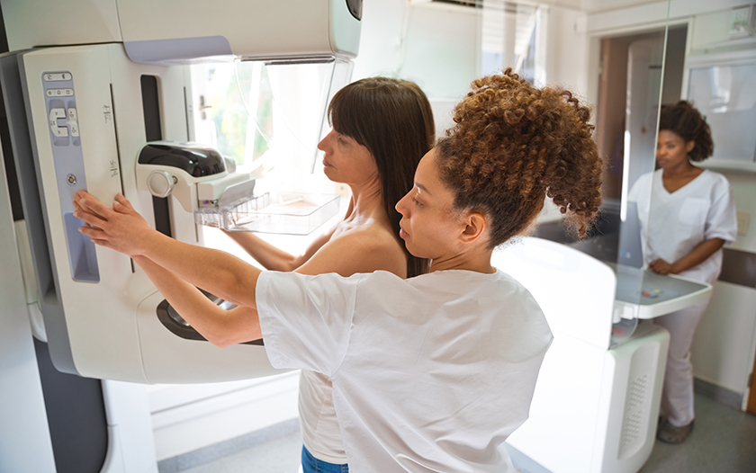
3D Mammography
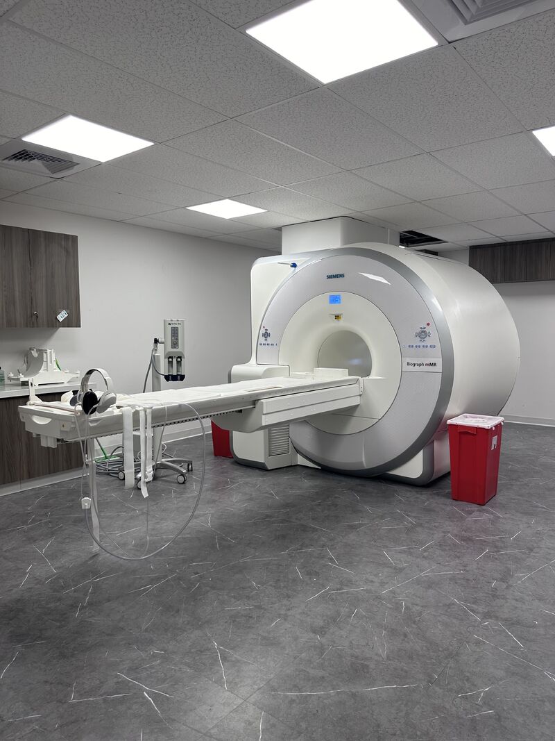
A 3D mammogram (also called breast tomosynthesis) is an imaging procedure that combines multiple breast X-Rays to create a three-dimensional picture of the breast. These multiple images of breast tissue slices taken with a 3D mammogram give radiologists a clearer image of the breast, making it easier to detect breast cancer. 3D mammography is especially beneficial for women with dense breast tissue.
Interventional Radiology
Interventional radiology is a specialization that uses image-guided, minimally invasive procedures to treat many conditions that once required traditional surgery. These treatments offer greater accuracy, less risk, less pain and a shorter recovery time than traditional surgical methods.
High-Field Open MRI
An MRI is an excellent tool for diagnosing and treating many health concerns. Using a magnetic field and radio waves, MRI is capable of viewing internal body parts such as the organs, tissues and bones. Most MRI machines are large, tube-shaped magnets and can be uncomfortable for many patients.
Open MRI machines however, provide for a more comfortable experience for patients that experience claustrophobia, have pain or mobility issues, or are larger in size. They are also an ideal solution for children.
PET/MRI
PET/MRI is a new, cutting-edge hybrid imaging modality that combines Positron Emission Tomography (PET) with Magnetic Resonance Imaging (MRI). This powerful diagnostic imaging tool allows providers to simultaneously evaluate the functional information provided by PET with the high-contrast anatomical information provided by MRI. PET/MRI has several clinical applications, most notably in Alzheimer’s, epilepsy and oncology imaging.
Arizona Diagnostic Radiology is a network of outpatient imaging centers located in Arizona. We believe the best way to prevent, detect and treat cancer involves both collaborative care and full patient participation.
View All Locations
