Breast Biopsy
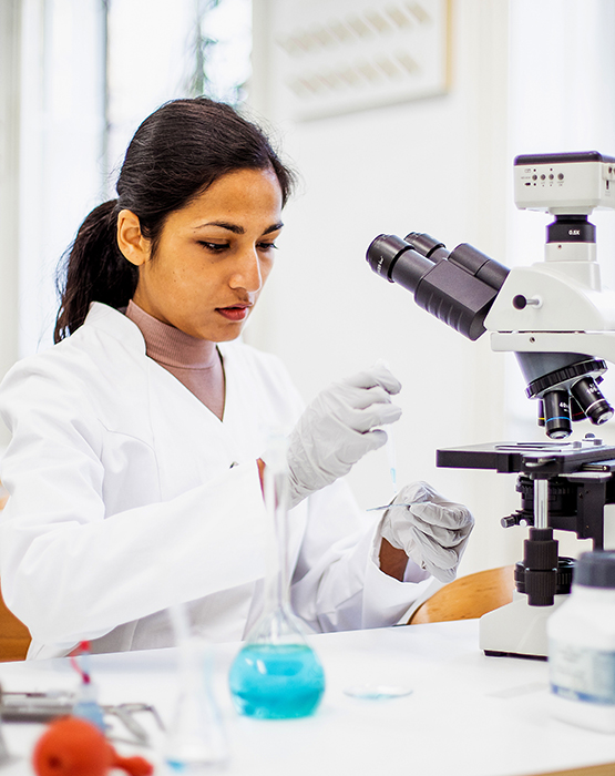
A breast biopsy is a procedure that removes a small sample of breast tissue for testing. The tissue sample is sent to a lab, where a pathologist (a physician who specializes in analyzing blood and body tissue) examines the tissue under the microscope and provides an analysis.
Casa Grande
1669 E McMurray Blvd, Casa Grande, AZ 85122 | (520) 876-0297
Gilbert Baseline
4915 E Baseline Rd, #116, Gilbert, AZ 85234 | (480) 354-9200
A breast biopsy is typically recommended when there is a suspicious area in the breast, such as a lump, or if there are unusual findings on a mammogram or breast ultrasound.
The results of a breast biopsy can show whether the area in question is breast cancer or if it is not cancerous. The pathology report from the biopsy can help your physician determine whether you need additional treatment.
Different techniques can be used to perform biopsy and the choice of procedure depends on each individual situation. A biopsy can be done by placing a needle through the skin into the breast to remove the tissue sample, or it can involve a minor surgical procedure.
At AZDRG, our fellowship-trained breast radiologists are proficient in performing various image-guided needle biopsies (if you require information on surgical biopsies, please consult with your physician).
Types of Biopsies
Image-guided biopsies include:
- Fine Needle Biopsy
Fine needle aspiration (FNA) is the least invasive method of biopsy and it usually leaves no scar. You will be lying down for this procedure and will be given an injection of local anesthesia to numb the breast. The radiologist uses a thin needle with a hollow center to remove a sample of cells from the suspicious area.
- Ultrasound-Guided Breast Biopsy/Stereotactic Needle Biopsy
In cases where an abnormality has been identified on imaging, the radiologist will use the appropriate imaging modality to guide a small caliber needle to the right location. When ultrasound is used, this is called “ultrasound-guided biopsy,” and if mammogram is used, it is called “stereotactic biopsy.”
- Core Needle Biopsy
This procedure is similar to FNA but the hollow needle that is used is larger. You will be lying down and your breast will be numbed with local anesthesia. Using the hollow needle, the radiologist will remove several cylinder-shaped samples of tissue from the suspicious area. In most cases, the needle is inserted about 3 to 6 times so that the doctor can get enough samples. Usually, core needle biopsy does not leave a scar.
Fine needle aspiration and core needle biopsy are both relatively quick procedures, taking about an hour to complete. They also both offer quick results without significant discomfort and scarring.


